Connect with Us
Looking for expert guidance on endoscopic solutions?
Reach out to us and explore imaging innovations that transform medical diagnostics.
Looking for expert guidance on endoscopic solutions?
Reach out to us and explore imaging innovations that transform medical diagnostics.


Products Sold
Installations
Endoscopy Centres
Patents
| OVP-SE II Plus | ||
|---|---|---|
| Image | 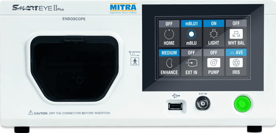 | |
| Model | OVP-SE II Plus | |
| Power Supply | Voltage | 100-240 VAC |
| Voltage fluctuation | Within ±10% | |
| Frequency | 50/60 Hz | |
| Frequency fluctuation | Within ±3 Hz | |
| Consumption electric power | 75VA | |
| Fuse rating | 4 A, 250 V | |
| Fuse size | Ø5.0 × 20.0 mm | |
| Size | Dimensions & Weight | 320(W) × 500(L) × 152(H) mm, 10.3kg |
| Classification (medical electrical equipment) | Type of protection against electric shock | Class I |
| Degree of protection against electric shock of applied part | Type BF applied part. Where no classification mark appears, the device is a TYPE BF applied part. | |
| Degree of protection against explosion | The Video Processor should be kept away from flammable gases. | |
| Observation | Video signal output | VBS composite (PAL), Y/C, HDMI, HD-SDI, DVI-D (Simultaneous output is possible.) |
| Video signal input | VBS composite (PAL), HDMI | |
| White balance | By pressing the white balance switch on the front panel, automatic white balance can be performed. | |
| Color tone adjustment | The following color tone adjustments may be using the color tone adjustment on the “System Setup” menu * “R” control:±10 steps * “B” control:±10 steps | |
| Iris area selection | The area over which light is measured can be changed according the body part that is being observed. The following to choices can be made using the iris mode selector on the front panel. Average: Normal observation Peak : When focusing on and/or observing a small bright area. | |
| Image enhancement mode setting | Structure enhancement in the endoscopic images can be enhanced electrically to increase the image sharpness. | |
| Gamma correction | Gamma correction adjustments can be performed by adjusting the gamma value. | |
| Switching the enhancement modes | The enhancement level can be selected from 3 levels enhancement modes (Low, Med. and High) using the image enhancement mode on the front button panel. | |
| Freeze screen display | The endoscopic image can be displayed as a frozen image, using the freeze switch key from endoscope. | |
| Brightness selector | The brightness can be adjusted using the brightness adjustment on the “system setup/camera adjustment” menu. | |
| Remote control | The following function can be controlled by the endoscope’s remote switches (Freeze, Record, Light, White Balance, mBLU, Enhancement, Contrast, Focus, Iris, Image Size, Remove Data & EXT In) | |
| mBLU | This mode is used to switch the normal mode to mBLU mode with white or blue light or vice versa. | |
| External Storage | USB Memory Stick | |
| Documentation | Patient Data | The following data and modes can be displayed on the video monitor using the keyboard - ID number, patient Name, Sex, Age, Date of Birth, Comments. |
| Display record state | Date/time (internal clock) | |
| Pre-procedural data | For a maximum of 40 patients- ID number, patient Name, Sex, Age, Date of Birth | |
| Image storage & retrieval | Memorization of selected setting | The following setting on the front panel are stored even when the video processor is turned OFF- System Setup, Setting the image record, Patient Data, Colour Tone, White Balance. |
| Memory Backup | Lithium Battery | 5-year life |
| Illumination | LED (White) | At Endoscope tip |
| Air Feeding Method | Pump | Diaphragm type pump |
| Airow | Low / High / Stop | |
| Airow Pressurization | Air pressurization of water container (detachable) | |
| OVP-SE II | ||
|---|---|---|
| Image | 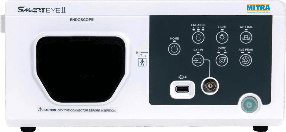 | |
| Model | OVP-SE II | |
| Power Supply | Voltage | 100-240 VAC |
| Voltage fluctuation | Within ±10% | |
| Frequency | 50/60 Hz | |
| Frequency fluctuation | Within ±3 Hz | |
| Consumption electric power | 75VA | |
| Fuse rating | 4 A, 250 V | |
| Fuse size | Ø5.0 × 20.0 mm | |
| Size | Dimensions & Weight | 320(W) × 500(L) × 152(H) mm, 10.3kg |
| Classification (medical electrical equipment) | Type of protection against electric shock | Class I |
| Degree of protection against electric shock of applied part | Type BF applied part. Where no classification mark appears, the device is a TYPE BF applied part. | |
| Degree of protection against explosion | The Video Processor should be kept away from flammable gases. | |
| Observation | Video signal output | VBS composite (PAL), Y/C, HDMI, HD-SDI, DVI-D Simultaneous output is possible. |
| Video signal input | VBS composite (PAL), HDMI | |
| White balance | By pressing the white balance switch on the front panel, automatic white balance can be performed. | |
| Color tone adjustment | The following color tone adjustments may be using the color tone adjustment on the “System Setup” menu * “R” control:±10 steps * “B” control:±10 steps | |
| Iris area selection | The area over which light is measured can be changed according the body part that is being observed. The following to choices can be made using the iris mode selector on the front panel. Average: Normal observation, Peak: When focusing on and/ or observing a small light area. | |
| Image enhancement mode setting | Structure enhancement in the endoscopic images can be enhanced electrically to increase the image sharpness. | |
| Gamma correction | Gamma correction adjustments can be performed by adjusting the gamma value. | |
| Switching the enhancement modes | The enhancement level can be selected from 3 levels enhancement modes (Low, Med. and High) using the image enhancement mode on the front button panel. | |
| Freeze screen display | The endoscopic image can be displayed as a frozen image, using the freeze switch key from endoscope. | |
| Brightness selector | The brightness can be adjusted using the brightness adjustment on the “system setup/camera adjustment” menu. | |
| Remote control | The following function can be controlled by the endoscope’s remote switches (Freeze, Record, Light, White Balance, Enhancement, Contrast, Zoom, Iris, Image Size, Remove Data & EXT In) | |
| mBLU | Switch to white or blue light | |
| External Storage | USB Memory Stick | |
| Documentation | Patient Data | The following data and modes can be displayed on the video monitor using the keyboard, ID number, Patient Name, Sex, Age, Date of Birth |
| Display record state | Date/time (internal clock) | |
| Pre-procedural data | For a maximum of 40 patients, ID number, Patient Name, Sex, Age, Date of Birth | |
| Image storage & retrieval | Memorization of selected setting | The following setting on the front panel are stored even when the video processor is turned OFF- System Setup, Setting the image record, Patient Data, Colour Tone, White Balance. |
| Memory Backup | Lithium Battery | 5-year life |
| Illumination | LED (White) | At Endoscope tip |
| Air Feeding Method | Pump | Diaphragm type |
| Airow | Low / High / Stop | |
| Water Feeding | Air pressurization of water container (detachable) | |
| OVP-S3 | |||
|---|---|---|---|
| Image | 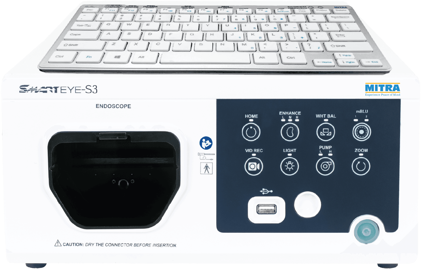 | ||
| Model | OVP-S3 | ||
| Power Supply | Voltage | 100-240 VAC | |
| Voltage fluctuation | Within ±10% | ||
| Frequency | 50/60 Hz | ||
| Frequency fluctuation | Within ±3 Hz | ||
| Consumption electric power | 18VA | ||
| Fuse | 3 A, 250 V Ø5.0 × 20.0 mm | ||
| Size | Dimensions & Weight | 445(W) × 320(L) × 152(H) mm, 8.9kg | |
| Classification | Type of protection against electric shock | Class I | |
| Degree of protection against electric shock of applied part | Depends on the applied part. (The degree of protection against electric shock of this product is BF type if the mounting part to be connected to this product is BF type. Note that CF type is not compatible with this product). | ||
| Degree of protection against explosion | The video processor should be kept away from flammable gases. | ||
| Observation | Analog SD TV signal output | Y/C x 1, VBS x 1 simultaneous output is possible. | |
| Digital signal output | HDMI x 2, DVI-D x 2 simultaneous output is possible. | ||
| White balance | By pressing the white balance switch on the front panel, automatic white balance can be performed. | ||
| Color tone adjustment | The following color tone adjustments may be done using the color tone adjustment on the “System Setup” menu. * “R” control: ±5 steps, * “G” control: ±5 steps, * ”B” control: ±5 steps, *Chroma: ±5 steps | ||
| Image settings | Contrast ± steps, Brightness: ± steps, Gamma: ±5 steps | ||
| Image size option | Small, Medium, Default & Large | ||
| Light target | ALC: ±5 steps | ||
| Iris area selection | The area over which light is measured can be changed according the body part that is being observed. The following two choices can be made using the iris mode selector on keyboard and endoscope remote switch. Average: Normal observation. Peak: When focusing on and/or observing a small bright area. | ||
| Image enhancement mode setting | Fine patterns or edges in the endoscopic images can be enhanced electrically to increase the image sharpness. | ||
| Gamma correction | Gamma correction adjustments can be performed by adjusting the gamma value. | ||
| Switching the enhancement modes | The enhancement level can be selected from 3 levels enhancement modes (Low, Med and High) using the image enhancement mode on the front button panel. | ||
| Brightness selector | The brightness can be adjusted using the brightness adjustment on the System & Image settings menu. | ||
| Contrast selector | The contrast can be adjusted using the contrast adjustment on the System & Image settings menu. | ||
| Zoom | 1.3x | ||
| Remote control | The following functions can be controlled by the endoscope remote switches (Light, White Balance, Freeze, Zoom, Record Video, Iris, Record Image, Enhancement, Image Size, mBLU, Remove Data, No Option) | ||
| *mBLU (Optional feature) | This mode is used to switch the normal mode to mBLU mode-1 & mBLU mode -2 | ||
| Freeze screen display | The endoscopic image can be displayed as a frozen image, using the freeze switch key from endoscope. | ||
| Pre-Freeze | While capturing an image, the function analyzes previous images & display the sharpest image from the view. | ||
| Portable Memory | Media | USB mass storage | |
| Documentation | Patient Data | The following data and modes can be displayed on the video monitor using the keyboard - ID number, Doctor Name, Patient Name, Sex, Age, Date of Birth, Comments | |
| Display record state | Date/time (internal clock) | ||
| Scope information data | The following scope information can be displayed on the monitor - Scope Model No., Serial No., Insertion Tube Diameter, Channel Diameter , Distal End Diameter, User details | ||
| USB Ports | Total 3 nos. | Keyboard & External Storage | |
| Recording format | Image | PNG | |
| Video | AVI | ||
| Memory backup | Number of user settings | User settings up to 5nos. users | |
| User function settings | Settings are held in video processor memory, even after video system is turned off. | ||
| Lithium battery | Life: 5 years | ||
| Illumination | Fiberless LED (White) | At Endoscope tip | |
| Air Feeding Method | Pump | Diaphragm type pump | |
| Airow | 3-mode (OFF, Low, High) | ||
| Water feeding | Method | Air pressurization of water container (detachable) | |
| Specifications | OVP-IN2 | OVP-EIN (New Series) | |
|---|---|---|---|
| Image | 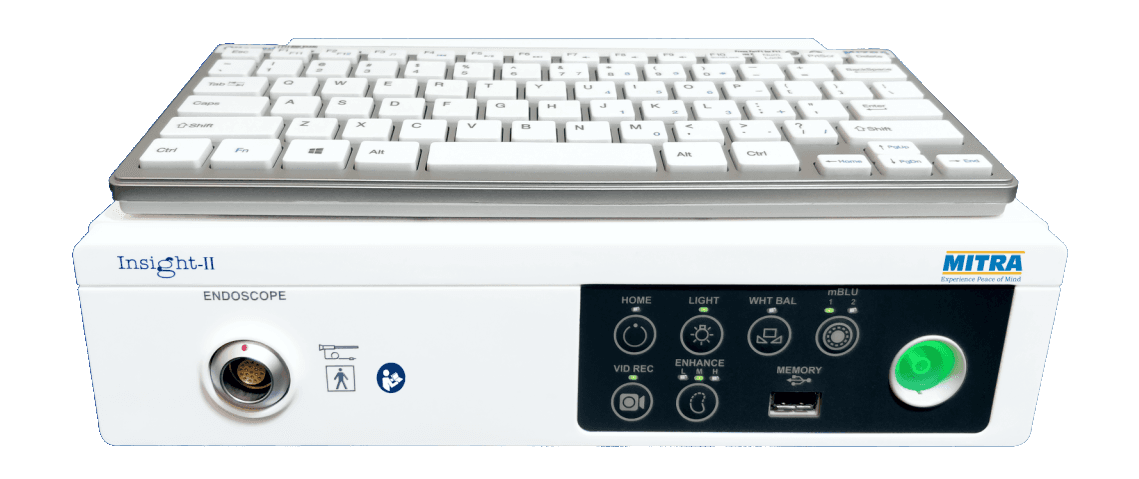 | 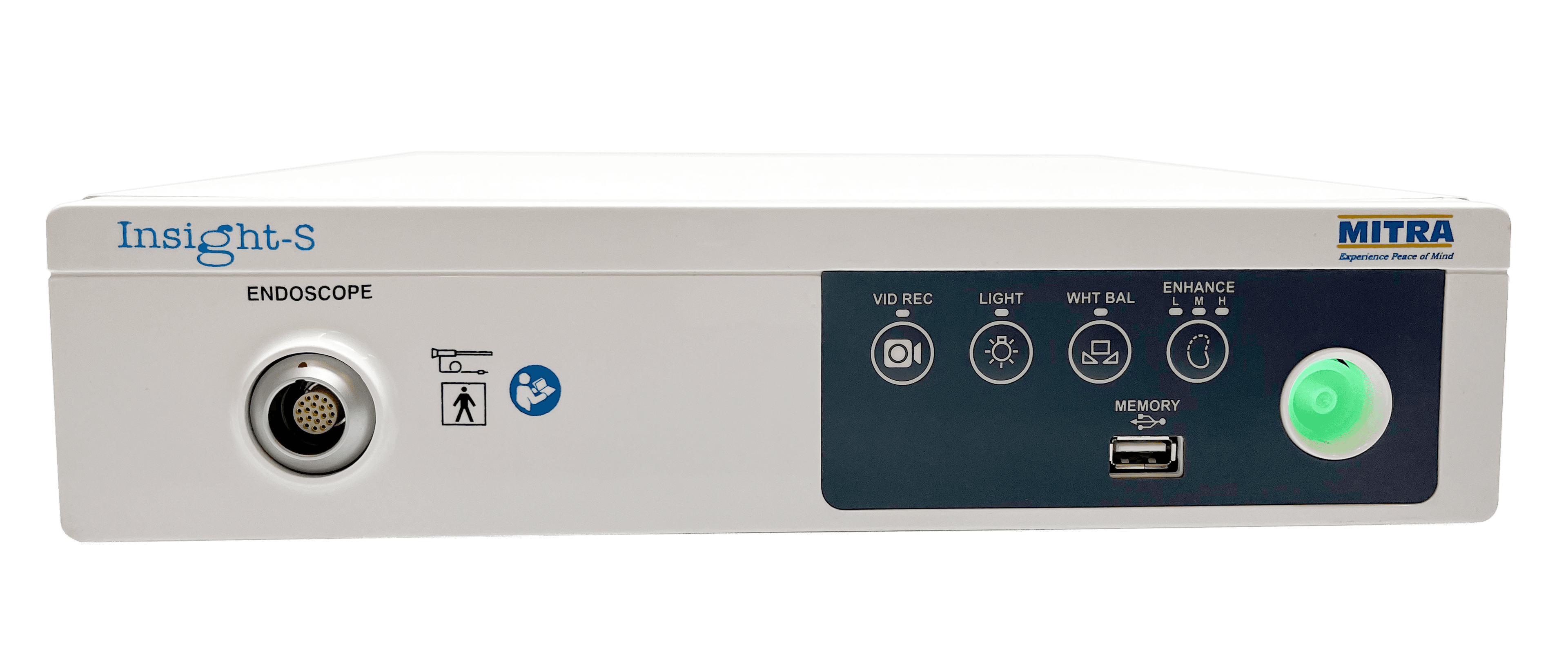 | |
| Power supply | |||
| Rated voltage | 100 – 240 V AC | 100 – 240 V AC | |
| Voltage fluctuation | Within ±10% | Within ±10% | |
| Rated frequency | 50/60 Hz | 50/60 Hz | |
| Frequency fluctuation | Within ±3 Hz | Within ±3 Hz | |
| Rated input | 18 VA | 18 VA | |
| Size | |||
| Dimensions | 295 (L) × 300 (W) × 80 (H) mm | 295 (L) × 300 (W) × 80 (H) mm | |
| Weight | 4.9 Kg Approx | 4.9 Kg Approx | |
| Classification (medical electrical equipment) | |||
| Type of protection against electric shock | Class I | Class I | |
| Degree of protection against electric shock of applied part | Depends on the applied part. (The degree of protection against electric shock of this product is BF type if the mounting part to be connected to this product is BF type. Note that CF type is not compatible with this product.) | ||
| Degree or protection against explosion | The video processor should be kept away from flammable gases. | ||
| Mode of operation | Continuous operation | Continuous operation | |
| Observation | |||
| Video signal output | HDMI x 2, DVI-D x 2, Y/C x 1, VBS x 1 simultaneous output is possible. | HDMI x 2, DVI-D x 2, Y/C x 1, VBS x 1 simultaneous output is possible. | |
| White balance | By pressing the white balance switch on the front panel, automatic white balance can be performed. | By pressing the white balance switch on the front panel, automatic white balance can be performed. | |
| Color tone adjustment | The following color tone adjustments may by using the color gain on the "System setup" menu *"R" control ±5 steps *"G" control ±5 steps *"B" control ±5 steps | NA | |
| Iris area selection | The area over which light is measure can be changed according the body part that is being observed. The following to choices can be made using the Iris mode selector on the keyboard. Average: Normal observation Peak: When focusing on and/or observing a small bright area. | NA | |
| Image enhancement mode setting | Fine patterns or edges in the endoscopic images can be enhance electrically to increase the image sharpness. | Fine patterns or edges in the endoscopic images can be enhance electrically to increase the image sharpness. | |
| Switching the enhancement modes | The enhancement level can be selected from 3 levels enhancement modes (Low, Med and High) using the image enhancement mode on the front button panel remote switch and keyboard. | The enhancement level can be selected from 3 levels enhancement modes (Low, Med and High) using the image enhancement mode on the front button panel. | |
| Gamma correction | Gamma correction adjustments can be performed by adjusting the gamma value. | NA | |
| Freeze screen display | The endoscopic image can be displayed as a frozen image, using the remote switch and keyboard. | The endoscopic image can be displayed as a frozen image, using the remote switch. | |
| Brightness selector | The brightness can be adjusted using the brightness adjustment on the "System & Image setting" menu. | NA | |
| Contrast selector | The contrast can be adjusted using the contrast adjustment on the "System & Image setting" menu. | NA | |
| Remote control | The following functions can be controlled by the endoscope's remote switches (Light, White Balance, Freeze, Zoom, Record Video, Iris, Record Image, Enhancement, Image Size, mBLU, Remove Data, No Option) | The following functions can be controlled by the endoscope's remote switches (Freeze, Image Record) | |
| mBLU | This mode is used to switch the normal mode to mBLU mode - 1 & mBLU mode - 2 | NA | |
| Documentation | |||
| Patient information | The following data can be displayed on the monitor: • Patient ID • Patient name • Gender • Age • Date of birth • Comment | NA | |
| Display the record state | Date/time (build in clock) | NA | |
| Pre-procedural patient data | The following patient data - ID number, Patient name, Sex, Age, Date of Birth. | NA | |
| Scope information data | The following scope information can be displayed on the monitor • Scope Model No. • Serial No. • Insertion Tube Diameter • Channel Diameter • Distal End Diameter • Cumulative uses • User details | NA | |
| USB ports | Keyboard & External Storage | 01 - External Storage | |
| Recording format | Image | PNG | PNG |
| Video | AVI | AVI | |
| Memory backup | Number of User Settings User settings | Up to 5Nos. users | NA |
| User Function Settings | Settings are held in video processor memory, even after video system is turned off. | NA | |
| Illumination | LED | LED at endoscope tip | LED at endoscope tip |